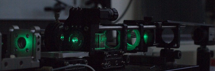

The Microfabrication Microsensing and Mechanobiology (3M) laboratory is based on a modern approach toward the controlled application and measurements of the forces developed within the living matter.
Several techniques developed by and for physicists, such as atomic force microscopy (AFM), optical forces manipulation and optical spectroscopy, nanopatterning and nanofabrication have been recently translated to the field of biology first and medicine then. In recent years, thanks to the application of AFM and optical tweezers (OT) to cells and biomolecules, such as the fabrication of mechanically engineered 2d and 3d scaffolds, the importance of the role of forces at a cellular and molecular level has been highlighted
The laboratory comprises several different shared facilities. A cell room co-managed with the animal house of the University of Trieste offer the standard environment for cell culture. Although most of the cell lines are developed through collaboration with universities across Europe, the cell room offers the possibility to develop these experiment autonomously. A bio-preparation lab, offers the basic equipment for the biological and biomolecular preparation. A section dedicated to prokaryotes is used for the preparation for microbiology experiments.
Four separated rooms host the analytical and microscopy instrumentation, that includes two AFMs, a micro-electro-mechanical system bench, , two optical tweezers microscopy (OTMs), one micro-Raman system, one digital holography microscopy (DHM). Several optical tables with different instrumentation as lasers and detectors are available to develop setups for specific applications, combining modules of microfluidics, optical microscopy and spectroscopy in a flexible manner.
The group participate to the management and operational activities of the FNF facility, for the micro and nano fabrication applied in different research field: sensors and detectors, from bio to high energy radiation. Electron and ion microscopy expertise are one of the strength of the group: a cross beam microscope is available for both, nanopatterning and deep analysis of samples. The development of the micromechanical, microfluidic and plasmonic tools for biophysical investigation is performed within the framework of FNF. The same framework is strongly involved in the develop of new architectures for next generation detector and sensor, in particular for FEL applcation, in operando and in situ devices for innovative studies with synchrotron in collaboration with IOM and Elettra beamlines.
The group is also actively involved in the Open-Lab project promoted by Area Science Park.
Instrumentation:
Local mechanical stimulation by using OTM and allowing to study cell mechanotransduction induced by piconewton forces.
Compact digital holography microscope (DHM): simple, portable optofluidic device for cell free-label analysis in liquid biopsy.
Real time generation of multiple holographic optical traps – for multiple cells mechanical stimulation
Synchronized nano-position control – optical trap scanning for OTM force measurement.
Development and fabrication of cantilevers for mechanical measurements on large cells and small multicellular bodies
Development of microfluidic platforms for cell sorting.
Several architectures for in operando characterization with synchrotron light.
New phosphors based screen for FEL beam diagnosys: PPD pixelated phosphor detector
SiN wires chips for FEL electron beam monitoring
Here we develop and apply several microscopy techniques to investigate in details the mechanics of several biological systems, ranging from mechanosensory neurons to cancer cells, from fertilization to heart failure, from bacteria to red blood cell infections, using a combination of microscopic, spectroscopic, and microfabrication techniques.
Many features of cell function such as motility, proliferation, differentiation and survival, can be altered by mechanical interaction (cell-cell, cell - ECM) even when the chemical environment remains unchanged. Controlled studies on cell mechanics and mechano-transduction are thus fundamental for developing new methodologies for diagnosis and therapeutic approaches in medicine, providing useful information for cell-disease phenotyping.
The same technical approach is applied in the field of detectors and sensors, in particular in the design and develop of new solution for emerging imaging and sensing technology. The application fields range from biosensing to Synchrotron and FEL beam monitoring, as a consequence of the strong interaction and collaboration with many beamlines of CNR and Elettra, and the electronics group of Elettra.