Facilities
Analytical Microscopy and Spectroscopy

STRAS

General informations

Location
TASC laboratory, Trieste, Italy
Main techniques and methods
Scanning Tunneling Microscopy (STM), Scanning Tunneling Spectroscopy (STS), Inelastic Electron Tunneling Spectroscopy (IETS), Low-Energy Electron Diffraction (LEED), Temperature Programmed Desorption (TPD)
Key instumentation
- Omicron Variable Temperature STM (140K-900K);
- Omicron Low Temperature STM (4K);
- FAST scan module compatible with both microscopes (up to 100 frames/s).

A scanning tunneling microscopy laboratory for the investigation of structural and electronic properties of solid surfaces at the nanoscale, with specific interest in describing their evolution during surface processes.

STRAS - Surface sTructure and Reactivity at the Atomic Scale

Technical description

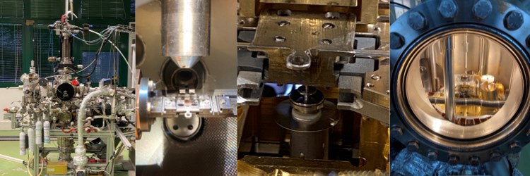
The STM laboratory includes two UHV systems equipped with a variable temperature Omicron VT-STM and a low temperature Omicron LT-STM.

The VT-STM has been modified in-house to allow for in-situ and in-operando measurements in the 140 – 900 K temperature range under exposure to reactive gases (O2, H2, CO, NO, etc.) at pressures up to 10-6 mbar. This system is ideal for the investigation of the kinetics of surface processes, such as elementary steps of catalytic reactions or growth and self-assembling, as well as for atomically resolved structural characterization of metal and oxide surfaces.

The LT-STM, for imaging at 4K, is capable of atomic scale resolution on weakly interacting adsorbates such as CO2 and organics of biological interest such as amino- and carboxyl- functionalized molecules. The LT system has been integrated with a home-built setup for single molecule vibrational spectroscopy measurements.

For a limited fraction of their activity (~10-15%), both systems are offered to external users through the NFFA-Europe program.

Development of new methods, instrumentation, software

Instrumentation: The technical capabilities of the laboratory have been further improved with the development of FAST, an add-on module that can upgrade virtually any scanning probe microscope to video rate imaging, allowing the investigation of dynamical processes at surfaces with unprecedented time resolution. FAST requires no modification of the existing scanner hardware, and its operation is totally transparent with respect to the existing electronics and acquisition software. The module, initially developed in collaboration with the Elettra Electronic Workshop group, has been improved in the recent years in collaboration with the Technical University of Munich and ICN2 in Barcelona. FAST can now routinely reach acquisition rates up to 100 frames/s while maintaining atomic resolution and its capabilities have been extended to atom-tracking and Fast-AFM. The user-friendliness of FAST was completed by the development of new acquisition and analysis softwares. FAST can now be purchased by contacting ILO-Elettra.

https://fastmodule.iom.cnr.it


Research Activity

STRAS - Surface sTructure and Reactivity at the Atomic Scale

The research activity of the group is dedicated to the study of surface structure and catalytic processes at the atomic scale. STM results are typically complemented by spectroscopic measurements and DFT calculations, both performed through external collaborations, inside and outside IOM.

In this context, two main research topics are presently addressed:

Growth, properties and reactivity of carbon-based 2D layers. Growth mechanisms for pristine and (N/B/TM-)doped graphene layers are investigated at the atomic scale during the growth process at technologically relevant temperatures and on both model surfaces and polycrystalline metal substrates, under hydrocarbon exposure and with time resolution up to video rate and above. Structural and electronic properties of the produced layer are investigated by VT-STM, LT-STM and STS, which are complemented by XPS, UPS and XPEEM/LEEM experiments. The reactivity of the layer is investigated by VT-STM under gas exposure. Experimental results are rationalized thanks to numerical simulations.

Self-assembling, structure and reactivity of organic layers. The systems of interest are thin films of organic semiconductor molecules (polyacenes, porphyrines, phthalocyanines) on transition metals or 2D materials. The assembling dynamics are investigated by means of both LT- and VT-STM. The morphology and the structure of the thin films are characterized by LT-STM at 4K and are complemented by electronic (STS) and vibrational (IETS) spectroscopy measurements at atomic/molecular level. STM/STS results are combined with RESPES, NEXAFS and SFG experiments and rationalized by numerical simulations (all of them performed through external collaborations).

Projects

  • H2020-INFRAIA-2018-2020, NEP, 2021-2026
  • PRIN 2017, MADAM, 2019-2022
  • H2020-INFRAIA-01, NFFA-Europe, 2015-2020

Main collaborations

  • University of Trieste, Italy
  • University of Milano Bicocca, Italy
  • Elettra Sincrotrone Trieste, Italy
  • Technische Universität Munich, Germany
  • Forschungszentrum Jülich, Germany
  • Technische Universität Dortmund, Germany

Key publications

Nanoscale, 112, 2022 doi:10.1039/d2nr02657k

Black or red phosphorus yields the same blue phosphorus film

Sala A., Caporali M., Serrano-Ruiz M., Armillotta F., Vesselli E., Genuzio F., Menteş T.O., Locatelli A., Comelli G., Africh C., Verdini A.
Nanoscale, 14-9:3589-3598, 2022 doi:10.1039/d1nr06485a

Carbide coating on nickel to enhance the stability of supported metal nanoclusters

Chesnyak V., Stavrić S., Panighel M., Comelli G., Peressi M., Africh C.
Advanced Functional Materials, 32-10, 2022 doi:10.1002/adfm.202105844

Quantum Confinement in Aligned Zigzag “Pseudo-Ribbons” Embedded in Graphene on Ni(100)

Sala A., Zou Z., Carnevali V., Panighel M., Genuzio F., Menteş T.O., Locatelli A., Cepek C., Peressi M., Comelli G., Africh C.
Carbon, 171:704-710, 2021 doi:10.1016/j.carbon.2020.09.056

“Inside out” growth method for high-quality nitrogen-doped graphene

Fiori S., Perilli D., Panighel M., Cepek C., Ugolotti A., Sala A., Liu H., Comelli G., Di Valentin C., Africh C.
Ultramicroscopy, 205:49-56, 2019 doi:10.1016/j.ultramic.2019.05.010

The new FAST module: A portable and transparent add-on module for time-resolved investigations with commercial scanning probe microscopes

Dri C., Panighel M., Tiemann D., Patera L.L., Troiano G., Fukamori Y., Knoller F., Lechner B.A.J., Cautero G., Giuressi D., Comelli G., Fraxedas J., Africh C., Esch F.
Nanoscale, 11-21:10358-10364, 2019 doi:10.1039/c9nr01072f

Doping of epitaxial graphene by direct incorporation of nickel adatoms

Carnevali V., Patera L.L., Prandini G., Jugovac M., Modesti S., Comelli G., Peressi M., Africh C.
Science, 359-6381:1243-1246, 2018 doi:10.1126/science.aan8782

Real-time imaging of adatom-promoted graphene growth on nickel

Patera L.L., Bianchini F., Africh C., Dri C., Soldano G., Mariscal M.M., Peressi M., Comelli G.
Carbon, 130:441-447, 2018 doi:10.1016/j.carbon.2018.01.010

Graphene on nickel (100) micrograins: Modulating the interface interaction by extended moiré superstructures

Zou Z., Carnevali V., Jugovac M., Patera L.L., Sala A., Panighel M., Cepek C., Soldano G., Mariscal M.M., Peressi M., Comelli G., Africh C.
Journal of Physical Chemistry Letters, 5-3:467-473, 2014 doi:10.1021/jz402609d

Atomic scale identification of coexisting graphene structures on Ni(111)

Bianchini F., Patera L.L., Peressi M., Africh C., Comelli G.
ACS Nano, 7-9:7901-7912, 2013 doi:10.1021/nn402927q

In situ observations of the atomistic mechanisms of Ni catalyzed low temperature graphene growth

Patera L.L., Africh C., Weatherup R.S., Blume R., Bhardwaj S., Castellarin-Cudia C., Knop-Gericke A., Schloegl R., Comelli G., Hofmann S., Cepek C.
Nature Chemistry, 4-3:215-220, 2012 doi:10.1038/nchem.1242

Controlling on-surface polymerization by hierarchical and substrate-directed growth

Lafferentz L., Eberhardt V., Dri C., Africh C., Comelli G., Esch F., Hecht S., Grill L.
Science, 309-5735:752-755, 2005 doi:10.1126/science.1111568

Chemistry: Electron localization determines defect formation on ceria substrates

Esch F., Fabris S., Zhou L., Montini T., Africh C., Fornasiero P., Comelli G., Rosei R.